Conventional Tomography Uses Which of the Following Principles
Moves opposite the x-ray tube in a seesaw motion b. Every year brings new advances in CT.
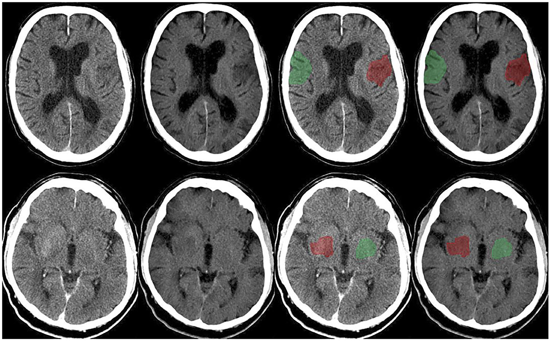
Frontiers Value Of Dual Energy Dual Layer Ct After Mechanical Recanalization For The Quantification Of Ischemic Brain Edema Neurology
Is focused to the automatic plane of interest d.

. 43 rows Tomography is imaging by sections or sectioning that uses any kind of penetrating wave. Learn faster with spaced repetition. The backscattered light is measured with an interferometric set-up to reconstruct the depth profile of the sample at the selected location.
Optical coherence tomography OCT A non-invasive optical diagnostic imaging technique which enables in vivo cross-sectional tomographic visualization of internal microstructure in biological systems. The principle of image reconstruction used in computed tomography can be utilized for the visualization of anatomical structures by the modulation of forms of energy other than x-radiation and it is also used in the field of nuclear medicine for the visualization of organs containing gamma- or positron-emitting radionuclides. For conventional radiography an x-ray beam is generated and passed through a patient to a piece of film or a radiation detector producing an image.
Is focused by the angle of movement c. During conventional tomography the image receptor. The principle advantage of CT imaging over other x-ray imaging is.
Hospitals outpatient clinics and physician offices find CT to be an essential tool for patient diagnosis and management. There are various specific and less particular indications for the use of conventional tomography but mostly it has been replaced by more efficient methods. C Conventional radiography cannot obtain sectional images.
Imaging Principles in Computed Tomography. Conventional tomography is now less commonly used because of the availability of cross-sectional imaging techniques such as US CT and MRI. You just studied 20 terms.
Tomography is an x-ray technique in which shadows of superimposed structures are blurred out by a moving x-ray tube. Uses of Conventional Radiography. If there is synchronous movement bw either of two among the three that is the patient the x-ray tube or the image receptor than there is blurring of image causes While movement only one thing is constant that is the fulcrum point of the tomographic equipment and the plane which posses this point is well demonstrated.
Examples include single-photon emission CT Single-photon emission CT SPECT Radionuclide scanning uses the radiation. Radiography is the most readily available imaging method. This technique may be useful in determining fracture healing evaluation of pulmonary nodules evaluating the kidneys in excretory urography and evaluating the integrity of spinal fusion.
Now up your study game with Learn mode. Optical coherence tomography. A useful analogy is to regard the technique as one that enables the patient to be imaged in slices like a loaf of sliced bread see Fig.
It typically uses light in the near-infrared spectral range which has a penetration depth of several hundred microns in tissue. The use of tomography makes possible the study of the trachea the bronchi and the blood vessels and the detection of infiltrates and cavities of the lungs calculi in the kidney gallbladder and bile ducts and tumors in the adrenals and urinary system. Conventional radiography involves the use of x-rays.
Although the principle of creating cross-sectional images is the same as for conventional CT whether single- or multi-slice the EBCT scanner does not require any moving parts to generate the. OCT is an optical imaging modality that is used to perform high resolution cross-sectional imaging of internal microstructures in materials and biological systems by measuring the echo time delay and magnitude of backscattered light. The method is used in radiology archaeology biology atmospheric science geophysics oceanography plasma physics materials science astrophysics quantum information and other areas of science.
Typically it is the first imaging method indicated to evaluate the extremities chest and sometimes the spine and abdomen. Basic Principle of Tomography. He originally applied reconstruction techniques in nuclear medicine ten years before he developed reconstruction techniques for computed tomography.
These same principles of tomographic imaging can also be applied to radionuclide scanning in which the sensors for emitted radiation encircle the patient and computer techniques convert the sensor data into tomographic images. OCT is analogous to ultrasound imaging except that it uses light rather than sound. Computed tomography CT has grown quickly from an innovative specialized tool to a mainstay of medicine in every healthcare setting.
Moves with the x-ray tube similarly to. The combined use of X-ray contrast media and tomography sectional bronchography. Examples include single-photon emission CT SPECT and positron-emission tomography PET.
Confocal scanning laser tomography See confocal scanning laser ophthalmoscope. There are 2 basic types of tomography. Study Tomography 1 flashcards from Thomas Couchs class online or in Brainscapes iPhone or Android app.
Each individual tomographic image or slice shows the. These same principles of tomographic imaging can also be applied to radionuclide scanning in which the sensors for emitted radiation encircle the patient and computer techniques convert the sensor data into tomographic images. Computed Tomography 4th Edition MULTIPLE CHOICE Sectional images are obtained from all of the following 1.
The term plain x-rays is sometimes used to distinguish x-rays used alone from x-rays combined with other techniques eg CT. Author not available 2003. Conventional tomography is a specialized radiographic technique developed originally for producing radiographs that showed only a section or slice of a patient.
The word tomography is derived from Ancient Greek τόμος tomos slice. It may be described as a radiograph obtained with a moving source image receptor assembly. These areas contain important structures with densities that differ from those of adjacent tissues.
Acquiring information from the patient by using special motions of the X-ray tube and detectors. The limitations of conventional tomography include all of the following except. Optical coherence tomography OCT is a non-invasive technique for cross-sectional tissue imaging.
2

X Ray Computed Tomography An Overview Sciencedirect Topics

Computed Tomography Of The Head An Overview Sciencedirect Topics

Customised Weight Based Volume Contrast Media Protocol In Ct Of Chest Abdomen And Pelvis Examination Journal Of Medical Imaging And Radiation Sciences

Frontiers Detecting Spurious Correlations With Sanity Tests For Artificial Intelligence Guided Radiology Systems Digital Health
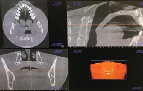
The Safe And Effective Use Of Cone Beam Computed Tomography

Pin On Medicine

Technological Developments Of X Ray Computed Tomography Over Half A Century User S Influence On Protocol Optimization European Journal Of Radiology

Dual Energy Ct Images Pearls And Pitfalls Radiographics
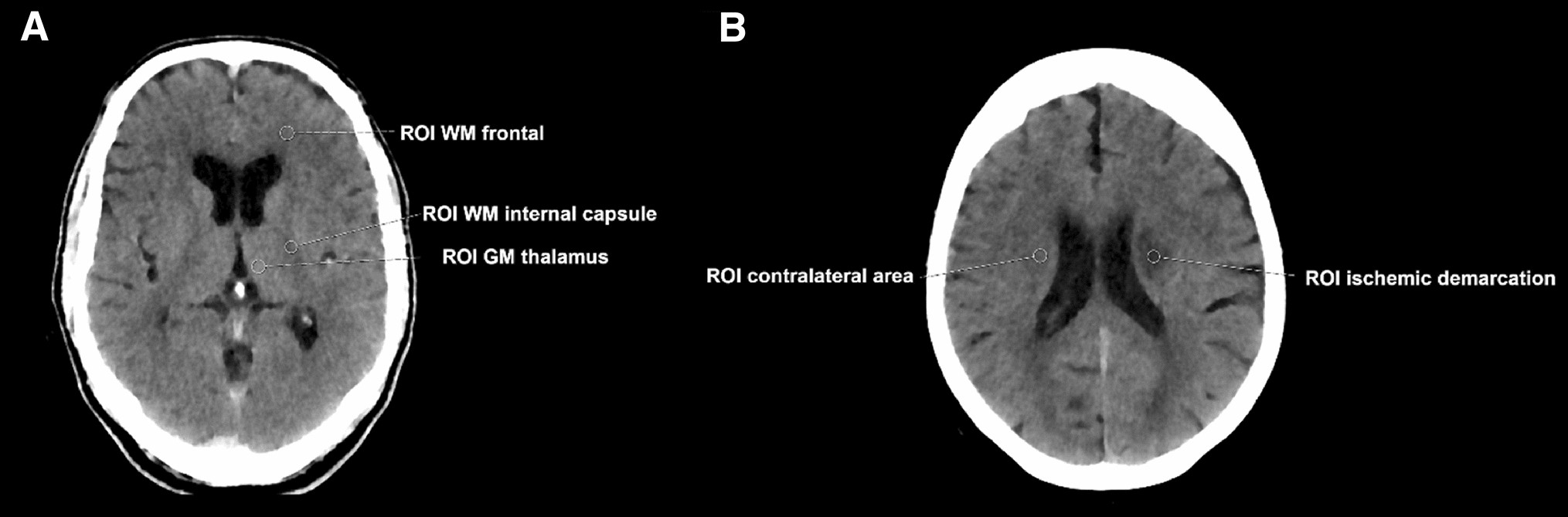
Impact Of Dose Reduction And Iterative Model Reconstruction On Multi Detector Ct Imaging Of The Brain In Patients With Suspected Ischemic Stroke Scientific Reports

Dual Energy Computed Tomography Radiologic Clinics

Low Dose Contrast Ct For Transcatheter Aortic Valve Replacement Assessment Results From The Prospective Spectacular Study Spectral Ct Assessment Prior To Tavr Journal Of Cardiovascular Computed Tomography
Linear Tomography Technology Britannica

What Is Tomography Youtube

What Is The Difference Between An X Ray A Ct Scan And An Mri

The Reveal Device Is An Implantable Loop Recorder Device That Wirelessly Records The Cardiac Rhythm It Is Different To A Usb Cardiac Rhythms Radiology Medical
Technological Developments Of X Ray Computed Tomography Over Half A Century User S Influence On Protocol Optimization European Journal Of Radiology

Single Photon Emission Computed Tomography Computed Tomography An Overview Sciencedirect Topics

Normal Kidneys On 4 Phase Ct Study Radiology Case Radiopaedia Org Radiology Study Case


Comments
Post a Comment